
| Year : 2007 | Volume : 1 | Issue : 2 | Page : 51-57 |    |
||
|

Purpose: The purpose of the present study was to analyze the effect of aging on activation capacity of shoulder muscles, between individuals of young and older age groups, during dynamic activities of pulling, pushing, elevation and throwing activities.
Materials and Methods: Nine young male adults of 20-29 year age group and eight old male individuals of 50-59 year age group, without any shoulder problems, were studied. Signals were recorded by surface electromyography as “percentage maximum voluntary isometric contraction” from middle deltoid, posterior deltoid, supraspinatus, and infraspinatus muscles of the shoulder. The results obtained from the muscles of younger age group were compared with those of older age group. Unpaired ‘t’ – test was used for purpose of statistical analysis.
Results: Significant increase in muscle activity was observed in elderly group as compared to younger group in activities involving pull, push, elevation and throw. Posterior deltoid (P P P P P P P P P P Conslusion: Greater recruitment of the selected shoulder muscles occurs, with aging, during the dynamic activity. With aging, the percentage of muscle fibers decrease, resulting in the decrease of muscle strength. This may imply that, as a compensatory mechanism, elderly individuals require greater recruitment of muscle fibers to perform an activity than younger individuals.
Clinical Relevance: Difference in recruitment pattern in the muscles of stability and mobility, between old and young individuals may be regarded as a potential factor for development of shoulder instability syndrome in elderly individuals.
Keywords: Aging, dynamic contraction, electromyography, shoulder muscles
| How to cite this article: Gaur DK, Shenoy S, Sandhu JS. Effect of aging on activation of shoulder muscles during dynamic activities: An electromyographic analysis. Int J Shoulder Surg 2007;1:51-7 |
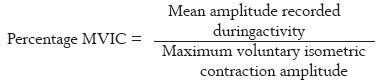
Electromyography (EMG) as a tool for the study of muscle function has been in use since the pioneering work of Inman et al . [1] Surface electromyography (sEMG) has been used to quantify muscle activity patterns and to analyze activation of shoulder muscles during sports-related activity [2] and routine day work. [3] An EMG variable like maximum voluntary contraction (MVC) is widely used to study the myoelectrical signals from the muscles. The use of MVC as a reference contraction is based on the idea that the amount of force produced varies directly with the myoelectrical output during signal recording. Thus, during MVC performed by an individual, myoelectrical signal is recorded in the form of MVC amplitude as ‘µVs’. For the purpose of comparison between different subjects, normalization is done. The mean amplitude of electrical activity recorded from the muscles during the contraction is divided by MVC amplitude and percentage MVC is obtained.
Although local muscle fatigue has been extensively studied in adults, [4],[5],[6] only few experiments have been available addressing differences or similarities in the neuro-muscular response, between young adults and older individuals, during static or dynamic contractions. [7],[8] Loss of muscle strength as a result of normal aging is reported to impair the functional ability in various communities. [9] This decline in strength is related to various normal aging processes, type of lifestyle and level of physical activity.
During any muscle contraction, according to size principle of motor unit (M.U.) recruitment, small type – I M.U.s are recruited first, followed by large type – II M.U.s.[10] Merletti
et al. [8] studied the effect of age on surface myoelectrical signals in healthy adults and older individuals and found that when voluntary contraction level was increased from 20% MVC to 80% MVC, the surface myoelectrical spectral signals increased; these were smaller in elderly subjects. This was explained on the basis that age-related decrease of number and size of fast twitch (type – II) fibers occurs, as indicated by histological data.
Although no relevant literature was available on the effect of age on MVC amplitude and %MVC of shoulder muscles recorded during dynamic activities of pull, push elevation and throw, a study [11] evaluated the influence of the aging process on properties of surface electrical and mechanical activity of biceps muscle during isometric contraction. The authors observed that, as compared to young controls, MVC was lower in the elderly subjects and root mean square and median frequency of EMG signals increased as a function of the effort level in both groups. Muscle activation pattern of four dynamic activities was evaluated by Illyes et al. [12] and it was found that deltoid and rotator cuff muscles of the recreational players showed stronger activity than professional players during pull, push, and elevation; during throwing, deltoid and rotator cuff muscles of professional players showed stronger activity.
The functions of shoulder muscles during various activities have been widely studied by EMG, and these provide a basis for designing rehabilitation programs for shoulder muscles. [13],[14],[15],[16] Definition of activation patterns of various shoulder muscles during dynamic activities may help in planning the rehabilitation process, as the accurate stimulation and training program of the injured and/or affected muscle can properly be planned, and regularly and accurately followed-up with EMG measurements. Earlier EMG studies indicated that the subscapularis muscle is important for anterior stability and infraspinatus for posterior stability. The infrapinatus muscle is active in abduction and flexion, and supraspinatus muscle in extension. [17],[18] The musculature of shoulder joint can be divided into intrinsic (stabilizing) muscles (supraspinatus, infraspinatus, biceps, and triceps) and extrinsic (mobilizing) muscles (anterior, middle and posterior deltoid, pectoralis major, latissimus dossi, teres minor). [19] Also, stabilizer muscles of the shoulder joint can be divided into high level stabilizers (supraspinatus, infraspinatus) and low level stabilizers (deltoid, biceps). [20] On the basis of the function, four shoulder muscles viz., middle deltoid, posterior deltoid, supraspinatus and infraspinatus, were selected for the study; the purpose of the study was to determine the difference in activation pattern of selected shoulder muscles, between young adults and older individuals, during dynamic muscles activities, using surface electromyography.
Subjects
Seventeen healthy male subjects participated in this study. All recordings were taken from the dominant side of the upper limb for each subject. Two groups were made for the purpose of comparison.
Group 1 involved nine subjects (sedentary young adults) between 20-29 years of age (Mean age, 24.11±1.66 yrs; mean weight, 65.38±9.08 kg; mean height, 170.94±5.62 cm). The physical activity level was level 4, i.e., no sports participation but can perform activities of daily living without any problem (Cincinnati sports activity scale, Barber-Westin et al. , 1999 [21] ). Random samples were drawn from students of different departments of GNDU, Amritsar.
Group 2 involved eight subjects (sedentary older individuals) between 50-59 years of age (Mean age, 56.5±3.10 yrs; mean weight, 74.62±13.52 kg; mean height, 173.12±6.49 cm). The physical activity level was level 4, i.e., no sports participation but can perform activities of daily living without any problem (Cincinnati sports activity scale , Barber-Westin et al. , [21] ). Random samples were drawn from staff members of different departments of GNDU, Amritsar.
Complete details of present and previous history, sports participation information and training characteristics were recorded for each subject. All subjects were screened for musculo-skeletal pain or disorders of the upper extremity and were excluded if they reported any present or previous disorder or symptom related to shoulder within past 6-12months. Each subject provided an informed consent prior to participation in the study approved by University Ethical Committee.
Procedure and instrumentation
Four pairs of disposable Ag-AgCl surface electrodes, 8 mm in diameter, with an inter-electrode distance of 2.5cm, were used for the study. The electrodes were attached to the skin over the muscle belly in the direction of muscle fibers. The reference electrode was taped to the seventh cervical spinous process. Recording electrodes were placed using the recommendation of Hermes et al . [22] European recommendations for surface EMG, results of SENIAM project were used (Middle deltoid (M.D) – at one-fourth the distance from the acromion process on the line joining acromion to lateral epicondyle of elbow; Posterior deltoid (P.D) – an area about two finger breaths behind the angle of acromion; Supraspinatus (SUP) – just rostral to the mid-point of the scapular spine and Infraspinatus (INF) – just below the mid point of the scapular spine.
The instrumentation for research included NORAXON 4 channel EMG, USA manufactured, with Myosytem 1200. The EMG signals were amplified by the amplifier system Driver Linx with the input impedance of 10-milli ohm. The following settings were used: bandwidth = 10 Hz – 500 Hz, common mode rejection ratio = 110 dB, sampling rate= 1000 Hz, RMS window = 100ms. The root mean square (RMS) values of EMG signals were calculated for consecutive segments of 50 ms. In order to allow comparison of the activity in specific muscles and the activity in specific muscles among different individuals, the EMG was normalized. The RMS values of EMG signals were calculated for each muscle by using maximum of three peaks EMG signals to represent 100% MVIC. Muscle activity was categorized as minimum (0-40%), moderate (40-70%) and maximum (70-100%).
MVC of all muscles were recorded in manual muscle testing position (MMT) in accordance with standard physical therapy guidelines, Daniel and Worthingm. [23] The subject was in sitting position on a stable base during all the recordings.
The MMT positions used were as follows: Middle deltoid: Arm abducted to 90º and elbow slightly flexed. Subject was asked to perform abduction with maximum force. Resistance to the movement was provided so that an isometric contraction resulted.Posterior deltoid: Arm in 90º of abduction and elbow flexed partially. Subject was asked to do extension. Resistance to the movement was provided so that an isometric contraction resulted.Supraspinatus: Arm in elevated position halfway between flexion and abduction (30º-45º anterior to coronal plane – scapular scaption) and shoulder internally rotated with thumb pointing down. The subjects were asked to do elevation in the same plane. Resistance to the movement was provided so that an isometric contraction resulted.Infraspinatus: Arm by the side of trunk and elbow flexed 90º. The subject was asked to perform lateral rotation. Resistance to the movement was provided so that an isometric contraction resulted.Three trials of MVCs were taken for the selected shoulder muscles for each subject and each muscle. During the recording of MVCs the subjects applied maximum force in the MMT position and were made to hold it for 5 sec and EMG activity was recorded simultaneously. The myoelectrical signals were recorded in the form of MVC amplitude as ‘µVs’. The best of the 3 trials was taken as the MVC. Thus for each subject one MVC for each muscle was recorded.
Four dynamic movements were investigated in the sequence of Pull, Push, Elevation and Throw. Exercises of pull, push and elevation involved the use of elastic green color heavy resistance theraband, 1.8 m. This theraband provided 5.0lb of resistance to stretch a 12 inch band length to 24 inches. [24],[25] In this study a 24 inch band was looped once so the total resistance provided was 10.0lb to stretch the band by 100%, i.e., 12 inches. The theraband was fixed to the wall at a height of 2feet. The same green color theraband was used for each subject while recording the EMG signals during the activities of pull, push and elevation. Hence a relatively constant force was provided during the dynamic movement by the theraband for both the groups studied. Each phase of pull, push and elevation exercise was performed at 40 beats per min, 0.1 sec beep duration, standardized with the aid of a metronome. Subjects were standing during all the activities. Each movement was repeated at least three times, one after the other.
The movements were performed as follows:
PULL [Figure – 1] The position of the subject was, standing at a feet distance from the wall, with chest facing the wall, holding the green color theraband in hand in starting position. The starting position of pull was in saggital plane with the arm at approximately 90º of flexion, elbow fully extended and forearm supinated 90º. From this position all subjects performed pull against the resistance administered by the green color heavy resistance theraband. The end point of pull was at 10º of extension at shoulder with 100º of flexion at elbow and 90º of supination at forearm.
Activity 1: Beep 1: gaining end position of pull with hold (1 sec) → Beep 2: Returning to starting position.
PUSH [Figure – 2] The position of the subject was, standing at a feet distance from the wall with back facing the wall holding the green color theraband in hand in starting position. The starting position of push was in saggital plane with the upper arm in neutral position beside the trunk with elbow in 90º of flexion, forearm in 90º of pronation and wrist in 30º of dorsiflexion. The end point of pushing was at 90º of flexion at shoulder with elbow at full extension and forearm and wrist remaining in the same position.
Activity 2: Beep 1: gaining end position of push with hold (1 sec) → Beep 2: Returning to starting position.
ELEVATION [Figure – 3] The position of the subject was, standing at a feet distance from the wall, with his side facing the wall, holding the green color theraband in hand in the plane of elevation. The plane of elevation was in approximately 20º flexion of frontal plane. The elbow and wrist were extended during the movement. Elevation started from a neutral position of the upper extremity. The end point was at approximately 120º of elevation.
Activity 3: Beep 1: gaining end position of elevation with hold (1sec) → Beep 2: Returning to starting position.
THROW [Figure – 4] The throw was performed with maximum speed and subjects were asked to throw as fast as they could, in the position that was natural for them. The starting position of throwing was shoulder at 90º of abduction and full external rotation with slight extension and elbow 90º flexed. A tennis ball, held in hand, was used for overhead throw with the target 5 m away into a net.
For the purpose of comparison between different subjects, normalization was done. The mean amplitude of electrical activity was recorded from the muscles during the dynamic activities of pull, push, elevation and throwing in ‘µVs’, separately for each activity, each muscle studied and each subject. This was then divided by MVC amplitude obtained for that particular muscle recorded in that individual subject and a “percentage MVC” was obtained, separately for each activity, each muscle studied and each subject.Percentage MVIC was calculated using the following formula:
Statistical analysis
Before statistical analysis, all EMG data was normalized to amplitude obtained during MVC and was taken in terms of %MVC. The significance level was calculated and corresponding level of confidence were determined by noting the P -values at 0.05 level of confidence. One-way ANOVA test was used for comparison between four muscles during an activity within the same group and mean values were used for interpreting the results obtained. Unrelated ‘t’ -test was used for comparison in the four muscles during an activity between the two different groups.
The mean values of %MVC (standard deviation), grading of activity of each muscle and significant difference between the two groups are summarized in [Table – 1].
Pull
In the sedentary adult group, all the four muscles demonstrated similar activation capacity with each muscle minimally active. However in sedentary old group only M.D and P.D were having similar activity, which was greater than SUP but the difference, was found to be statistically insignificant.
Comparison between the adult and old groups showed statistically significant difference in P.D (t=2.007, P P
PushDuring pushing, minimal activity was obtained in all four muscles in both groups. M.D and INF, and P.D and SUP muscles showed similar activity in both groups, with greatest activity in M.D in young group and in INF in old group.
Comparison between the groups showed statistically significant difference in M. D (t = 2.22, P < 0.02), SUP (t = 4.16, P P
ElevationDuring elevation, M.D in both young and old groups exhibited maximal activity while others were moderately active. Similar activation capacity was found in P.D, SUP and INF with M.D mainly taking part in the movement.
Comparison between the groups exhibited significant difference in P.D (t = 1.56, P < 0.05) and INF (t = 1.66, P
ThrowIn the young group moderate activity was obtained in SUP while others were minimally active. In the old group all muscles were found to be maximally active. SUP demonstrated maximal activity in young group while the INF in old group.
Comparison between the groups depicted significant difference in M.D (t = 1.97, P P < 0.009) activity with greater activation in the old group for M.D, P.D and INF. In the present study, four shoulder muscles were investigated; the purpose was to analyse the difference in muscle activation patterns among healthy young adults and older individuals, during dynamic activities of pull, push, elevation and throw. Surface electromyography was performed to obtain the data, and the bandwidth of the amplifier was sufficient for dynamic motion recordings. For data processing, “percentage MVC” of each muscle was used to compare various muscle activities in different subjects during movements.Though the actual amount of the force generated over a range was not measured by using dynamometer during the activities, an attempt to keep the force output constant (by use of constant resistance using green color theraband) was made. This implied that to pull, push and elevate through a constant distance all subjects had to exert a constant force, however the amount of motor units recruited to produce this force would vary between groups of subjects studied. This was measured with sEMG.
The comparisons made in this study showed that %MVC obtained for selected shoulder muscles in the study were greater in elderly individuals for all four dynamic activities [Table – 1]. During pulling, greater activity was found in posterior deltoid and infraspinatus. During pushing middle deltoid, suprapinatus and infrapinatus exhibited greater activity. During elevation greater activity was obtained in posterior deltoid and infraspinatus again. Lastly during throwing greater activity occurred in middle deltoid and infraspinatus. Hence greater muscle activation occurred in older individuals with increase in the effort for activities from pulling to throwing. This higher activity may be due to variability of motor unit discharge rate at high force levels. [26] Since older individuals exhibit reduced force control and have slower neuro-muscular contractile properties, greater proportion of motor unit recruitment may occur in older individuals during a high effort activity in order to compensate for decrease in muscle strength that occurs with age. As quantified from this study, 4.6%-18.9% greater activation of stabilizer muscles (supraspinatus and infraspinatus), and 2.7%-19% greater activation of mobilizer muscles (middle deltoid and posterior deltoid) occurs in older individuals during dynamic activity [Table – 1].
During pushing and throwing the primary stabilizer muscle of shoulder joint, i.e, infraspinatus, was more active, and during pulling and elevation, mobilizer muscles, i.e., posterior deltoid and middle deltoid were more active in older individuals as compared to young adults. Anders [27] and Cain [28] had similar findings that infraspinatus muscle was most effective in providing dynamic stability in abduction and throwing, acting as high level stabilizer. Thus it can be observed from this study that aging alters the activation pattern of the muscles during dynamic activities.
Therefore the muscle activity patterns obtained may have clinical implications for both the training and maintenance of healthy muscle function in old age as follows:
- The study may help in providing basis for designing exercise programs for veteran individuals based on the pattern of muscle recruitment during different movements at the shoulder joint.
- By recognizing the different patterns of activity of mobilizer and stabilizer of shoulder, key muscles may be trained for relevant activity or task. This may thus reduce the risk of injury and have a role in injury prevention among older age group.
- It has been concluded by De Serres [29] that decline in strength with age, does not appear to result from an impairment of the neural drive to muscle and might be related to loss of muscle mass and decreased physical activity. Also based on the results of this study, it is recommended that in order to reduce the loss of muscle mass that occurs as an individual ages, a program of ‘active ageing’ which includes dynamic resistance training for rotator cuff or shoulder muscles should be incorporated.
Hence it can be concluded from the study that, with aging, elderly individuals require greater recruitment of muscle fibers in performing any shoulder activity than younger adults to compensate for loss in muscle strength. Also in sedentary old individuals, a stabilizer muscle like infraspinatus is active to a greater extent during dynamic muscle activity like pushing and throwing, as compared to young adults.
| 1. | Inman VT, Saunders JB, Abbott LC. Observations on the function of the shoulder joint, 1944. Clin Orthop Relat Res 1996;330:3-12.  [PUBMED] [FULLTEXT] [PUBMED] [FULLTEXT] |
| 2. | Heise GD. Electromyography changes in agonist muscle during practice of a multi-joint throwing skill. J Electromyogr Kinesiol 1995;5:81-94. |
| 3. | Schuldt K, Ekholm J, Harms-Ringdahl K, Nemeth AG. Influence of sitting postures on neck and shoulder electromyography during arm-hand work movement. Clin Biomech 1987;2:126. |
| 4. | Bigland-Ritchie B, Donovan EF, Roussos CS. Conduction velocity and EMG power spectrum changes in fatigue of sustained maximal efforts. J Appl Physiol 1981;51:1300-5. [PUBMED] [FULLTEXT] |
| 5. | Moritani T, Muro M, Nagat A. Intramuscular and surface electromyogram changes during muscle fatigue. J Appl Physiol 1986;60:1179-85. |
| 6. | Bilodeau M, Erb MD, Nichols JM, Joiner KL, Weeks JB. Fatigue of elbow flexor muscles in younger and older adults. Muscle Nerve 2000;24:98-106. |
| 7. | Yamada H, Okada M, Oda T, Nemeto S, Shiozaki T, Kizuka T, et al . Effects of aging on electromyography variables during fatiguing isometric contractions. J Hum Ergol Tokyo 2000;29:7-14. |
| 8. | Merletti R, Lo Conte LR, Cisari C, Actis MV. Age related changes in surface myoelectric signals. Scand J Rehabil Med 1992;24:25-36. [PUBMED] |
| 9. | Vandervoort AA, McComas AJ. Contractile changes in opposing muscles of the human ankle joint with aging. J Appl Physiol 1987;61:361-7. |
| 10. | Halin R, Germain P, Bercier S, Kapitaniak B, Buttelli O. Neuro-muscular response of young boys versus men during sustained maximal contraction. Med Sc Sp Ex 2003;3:1042-8. |
| 11. | Esposito F, Malgrati D, Veicsteinas A, Orizio C. Time and frequency domain analysis of electromyogram and sound myogram in the elderly. Eur J Appl Physiol Occup Physiol 1996;73:503-10. [PUBMED] |
| 12. | Illyes A, Kiss RM, Shoulder muscle activity during pulling, pushing, elevation and overhead throw. J Electromyography Kinesiol 2005;15:282-9. |
| 13. | Bradley JP, Tibone JE. Electromyographic analysis of muscle action about the shoulder. Clin Sports Med 1991;10:789-805. [PUBMED] |
| 14. | Kronberg M, Nemeth G, Brostrom LA. Muscle activity and coordination in the normal shoulder. Clin Orthop Relat Res 1990;257:76-85. |
| 15. | Moseley JB Jr, Jobe FW, Pink M, Perry J, Tibone J. EMG analysis of the scapular muscles during a shoulder rehabilitation program. Am J Sports Med 1992;20:128-34. |
| 16. | Townsend H, Jobe FW, Pink M, Perry J. Electromyographic analysis of the glenohumeral muscles during a baseball rehabilitation program. Am J Sports Med 1991;19:264-72. [PUBMED] |
| 17. | Hovelius L. Anterior dislocation of the shoulder. Phd Thesis. unpublished doctoral thesis. Link φping University No. 139. Link φping: Sweden; 1982. |
| 18. | Saha AK. Dynamic stability of the glenohumeral joint. Acta Othop Scand 1971;42:491-505. |
| 19. | Irlenbusch U. Muscles of the shoulder joint and functions. Thieme: New York; 1999. |
| 20. | Kelly BT, Backus SI, Warren RF, Williams RJ. Electromyographic analysis and phase definition of the overhead football throw. Am J Sport Med 2002;30:837-44. |
| 21. | Barber-Westin SD, Noyes FR, McCloskey JW. Rigorous statistical reliability, validity, and responsiveness testing of the Cincinnati knee rating system in 350 subjects with uninjured, injured, or anterior cruciate ligament-reconstructed knees. Am J Sports Med 1999;27:402-16. [PUBMED] [FULLTEXT] |
| 22. | Hermes HJ, Freriks B, Merletti R, Stegemann D, Blok J, Rau G, et al . European recommendations for surface EMG, results of SENIAM project, Roessingh research and development. B.V. Enschede: the Netherlands; 1999. |
| 23. | Daniel and Worthingm, Lucille. Muscle testing: Technique of manual examination, 3 rd edition. W B Saunders Company: 1972. |
| 24. | Available from: http://www.binderpendent.com/hompg/bidindep/store/aisles/exercise/bands/theraband.htm. |
| 25. | Available from: http://thera-bandacademy.com/elements/clients/docs/TBinstruction-012706_013106_130451.pdf. |
| 26. | Roos MR, Rice CL, Vandervoort AA. Age-related changes in motor unit function. Muscle Nerve 1997;20:679-90. [PUBMED] [FULLTEXT] |
| 27. | Anders C, Bretsehneiders S, Bernsdorf A, Erler K, Schneider W. Activation of shoulder muscles in healthy men and women under isometric conditions. J Electromyogr Kinesiol 2004;14:699-707. |
| 28. | Cain PR, Mutschler TA, Fu FH, Lee SK. Anterior stability of the glenohumeral joint: A dynamic model. Am J Sport Med 1987;15:144-8. |
| 29. | De Serres SJ, Enoka RM. Older adults can maximally activate the biceps brachii muscle by voluntary command. J Appl Physiol 1998;84:284-91. [PUBMED] [FULLTEXT] |
[Figure – 1], [Figure – 2], [Figure – 3], [Figure – 4]
[Table – 1]